
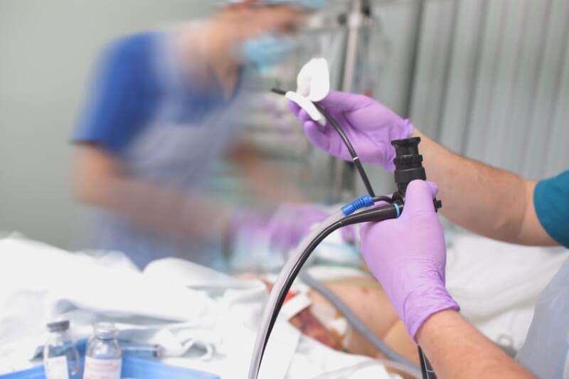
Endoscopy is a medical term that is used interchangeably with other procedures. In reality, endoscopy refers to a range of examinations that involve a camera called an endoscope.
So, what is the difference between an upper endoscopy and an endoscopy? An upper endoscopy refers to the endoscopy procedure concerning the upper GI tract. Its goal is to examine the esophagus, stomach, and duodenum. Another gastro endoscopy called the colonoscopy is performed to examine the lower GI tract, specifically the colon.
Knowing the difference between an endoscopy and upper endoscopy will help you understand what screening method is applicable to your symptoms.
What Is an Endoscopy?
An endoscopy is a procedure involving an endoscope, a flexible tube attached with a camera and a light that transmits live images onto a computer. Endoscopy is primarily a screening method used to diagnose and treat diseases. Other procedures can be performed alongside an endoscopy to complete a patient’s treatment or diagnosis procedure.
A Brief History
Historians believe that inventions similar to the endoscope were around as early as the ancient Roman and Greek periods. But it wasn’t until 1805 that a device specifically used to examine the gastrointestinal tract was invented. The device known as Lichtleiter or “light guiding device” was created by Phillip Bozzini to examine the pharynx, rectum, and urinary tract.
The word endoscope was first used and coined in 1853 when inventor Antoine Jean Desormeaux created a similar instrument specifically designed to examine the bladder and urinary tract.
Iterations of the same device have been introduced since then. In 1868, Dr. Adolph Kussmaul used the device to observe the inside of the human stomach, which was the first attempt to do so in human history. In 1881, Johann von Mikulicz and his peers created an endoscope similar to what we have today. However, the flexible iteration of this device only came around in 1932 that allowed stomach examination by using lens and a light to reflect the inside of a person’s stomach.
More modern innovations on the endoscopy began in the late 1900s, particularly the photographic gastronomic camera developed by Japanese doctors. New materials such as glass fiber allowed American inventors to find ways to examine the stomach, however, photographic limitations applied, rendering the new material irrelevant.
Finally, in 1964, a “gastronomic camera” was created, allowing doctors and surgeons to record stomach contents and view them as photographs. Eventually, gastronomic cameras were replaced by fiberscopes and this lead to the development of videoscopes.
With the advent of video cameras and TV display, doctors were able to enhance the process of endoscopy by projecting the live feed of the examination to HD screens. With modern endoscopy, nurses, doctors, and other medical professionals can study the stomach in real-time and make more accurate diagnoses thanks to high-quality resolution technology.
Purpose and Applications
1. Diagnosis
An endoscopy allows doctors to understand the nature of a disease. Additional procedures, such as a biopsy (the process of removing tissues for further study, usually by a pathologist), may be performed to rule out the possibility of diseases.
2. Screening and Investigation
Bleeding, pain, fever, heartburn, nausea, and vomiting can be a cause for concern. An endoscopy is recommended by doctors as a way of verifying the cause for these symptoms.
3. Treatment
An endoscopy can also be used for treatment purposes. Doctors can use the endoscope to study anatomical changes, especially for structural diseases. Using the camera, doctors can perform visual examination on a site and determine whether the current treatment plan is effective or not.
An endoscopy may also be performed to treat certain disorders. For instance, an upper GI endoscopy can be used to treat bleeding stomach ulcers.
Endoscopy VS Upper Endoscopy
Endoscopy describes a range of medical procedures that use an endoscope, which includes upper endoscopy. Upper endoscopy or esophagogastroduodenoscopy (EGD) refers to the endoscopic procedure studying the upper GI tract. The gastroenterologist inserts the tube through the mouth down to the small intestine to look for ulcers, growth, and other gastrointestinal abnormalities concerning the upper portion of the GI tract.
On the other hand, endoscopy refers to any screening procedure that uses a thin, flexible tube with a camera and light attachment. There are many types of endoscopy procedures available that involve other parts of the body.
Types of Endoscopy
Below are the various endoscopy procedures done depending on the area being examined, treated, or diagnosed:
1. Arthroscopy
Performed by making a small incision near a joint. An endoscope is passed through the joint to evaluate joint disorders such as arthritis. Arthroscopy is also used to repair joint tears and minor damages.
2. Bronchoscopy
Bronchoscopy is performed to study the bronchial tubes or the large tubes of the lungs branching into bronchus and bronchioles. This is done to look for tumors and growths in the lungs. Other medical procedures can be carried out alongside a bronchoscopy such as a biopsy or dilation.
3. Colonoscopy
Colonoscopy is one of the most common endoscopy procedures, routinely performed for patients 50 years old and above since they are at a higher risk of developing colon cancer. Patients undergoing colonoscopy are required to drink a colon prep drink to ensure accurate results. A colonoscopy is a procedure performed by inserting the endoscope through the rectum to look for colon polyps as a measure against colon cancer.
4. Colposcopy
Cervical cancer is often diagnosed through pap smear. Colposcopy is recommended after doctors find reasons for further investigation following a pap smear. During a colposcopy, an endoscope is inserted through the vagina to study the cervix for signs of cervical cancer.
5. Cystoscopy
Disorders concerning the bladder can be diagnosed through a procedure called cystoscopy. During the exam, a thin tube is inserted through the urethra (the long tube where urine is transported from the bladder) to detect early signs of bladder cancer, for instance.
6. Endoscopic Retrograde Cholangiopancreatography
Also known as ERCP, this type of endoscopy is inserted through the mouth down towards the pancreatic ducts. Unlike the upper GI, an ERCP exam is performed to study the pancreatic ducts in the liver and pancreas. The ERCP can be performed as a minimally invasive, non-surgical method of retrieving gallstones.
7. Laparoscopy
A laparoscopy is a known method for removing an appendix, as a treatment for appendicitis. This procedure is also useful in determining a host of disorders concerning organs in the abdominal region, including infertility and liver problems. Laparoscopic surgery refers to the minimally invasive surgery achieved by making a small incision (usually half an inch long) where the laparoscope (camera) is inserted to accomplish the surgery.
8. Laryngoscopy
Persistent coughing, throat pain, and bad breath are usually no cause for worry, but can be symptomatic of worse problems concerning the larynx. Laryngoscopy is performed to investigate these symptoms and visualize the larynx, often looking at growths in the throat or vocal cords to understand the cause of the problem.
9. Mediastinoscopy
Similar to an arthroscopy, mediastinoscopy is performed by making a small incision, this time above the breast bone. This procedure is used to examine the middle of the chest, also for lymph node removal in the case of lung cancer.
10. Proctoscopy
The rectoscope, the tool used during a proctoscopy, is a bit different from the usual endoscope. Instead of a long, flexible tube, the rectoscope is a straight hollow tube with a small light bulb at the end used to inspect minor rectal problems such as hemorrhoids or more serious ones such as a rectal polyp.
11. Upper GI Endoscopy
Also referred to as esophagogastroduodenoscopy (EGD), this procedure is done by inserting a thin tube in the mouth to observe the esophagus, stomach, and duodenum. During an upper endoscopy, doctors may elect to perform other procedures such as esophageal dilation as a treatment for a narrowed esophagus.
Learn more: A Complete Overview of Upper GI Endoscopy
Tools Used During Endoscopy
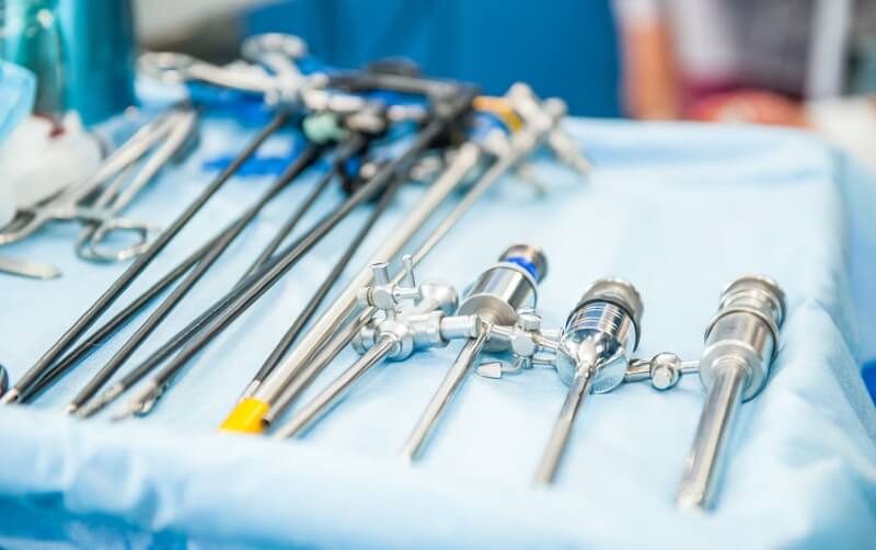
Aside from the endoscope, doctors use other tools to perform an exam. These include:
- Cytology brush for acquiring tissue and cell samples
- Flexible forceps used to carefully acquire tissue samples
- Suture removal tools that allow doctors to remove stitches inside the body
- Biopsy forceps designed to take samples of suspicious tissue and growth
Advancements in Endoscopy
Although modern endoscope has gone a long way from its early versions, contemporary inventors are still finding ways to improve the device. The current endoscope requires insertion through the mouth, anus, or by making a small incision in the treatment area. Although minimally invasive, this procedure can be off-putting for patients and could be detracting thousands of others who need to be screened.
Advancements in endoscopy exist to eliminate the pain points of current endoscopic procedures. Two note-worthy developments include:
Virtual Endoscopy
Virtual endoscopy works by using CT and MRI scans to reconstruct an area of the body. Three-dimensional pictures rendered in high quality are recreated to help doctors envision certain problem areas without the need for an endoscope.
Virtual endoscopy is currently being used to inspect the urinary tract. While it is yet to replace cystoscopy completely, virtual endoscopies allow for the detection of small bladder abnormalities including lesions less than 5 mm.
Virtual endoscopy is also being used as a competitive alternative to colonoscopy. With virtual endoscopy, no sedation is required and doctors are able to examine the entire colon even in patients with tumors. However, polyp screening efficiency tends to decrease with decreasing polyp size.
Capsule Endoscopy
Another non-invasive procedure, capsule endoscopy is a unique technology that involves tiny cameras placed inside a capsule, which is swallowed by the patient. As the capsule travels through the intestine, the camera takes multiple photos of the small intestine and transmits these photos on a data recorder worn by the patient around the waist.
Among its many benefits, capsule endoscopy allows clear visualization of the esophagus, stomach, bowels, and colon. It is also beneficial in detecting lesions and occult bleeding (bleeding that is invisible to the naked eye).
Patients are asked to return to the medical facility 8-10 hours after ingesting the camera to retrieve the recording device. The camera will be naturally flushed out through bowel movement.
Although rare, it’s possible for the tiny camera to get lodged in the small intestine, causing obstruction. When this happens, the capsule has to be removed through surgery or an upper endoscopy, depending on the location of the camera.
Schedule Your Endoscopy Today
At Gastro Center NJ, our facilities are fully equipped to perform gastrointestinal endoscopy.
Not sure what endoscopy procedure you should get for your symptoms? Get in touch with us today for a consultation.